Home
Uncategories
Anatomy Lateral Epicondyle Of Humerus - Lateral Epicondyle Fracture Adults : Lateral epicondyle (epicondylus lateralis) is a rounded projection at the distolateral end of the humerus.
Anatomy Lateral Epicondyle Of Humerus - Lateral Epicondyle Fracture Adults : Lateral epicondyle (epicondylus lateralis) is a rounded projection at the distolateral end of the humerus.
Anatomy Lateral Epicondyle Of Humerus - Lateral Epicondyle Fracture Adults : Lateral epicondyle (epicondylus lateralis) is a rounded projection at the distolateral end of the humerus.. The posterior surface of the lateral epicondyle serves as an attachment point for some of. The common extensor tendon attaches to the lateral epicondyle, acting as the common attachment for the superficial extensor muscles of the forearm. Yes, the most lateral epicondyle, the most lateral part of the humerus, can be felt through the skin. A common injury associated with the lateral epicondyle of the humerus is lateral epicondylitis also known as tennis elbow. Gross anatomy human anatomy anatomy and physiology occupational therapy massage muscles action group action.
Three nerves are directly related to the humerus and are, therefore, liable to injury: They serve as attachment points for the muscles of your lower arm, wrist, and hand. Lateral epicondylitis, also known as tennis elbow, is the most common overuse syndrome in the these muscles originate on the lateral epicondylar region of the distal humerus. The unity of form and function. The elbow is a hinge joint consisting of three bones:
Humerus Wikipedia from upload.wikimedia.org Your humerus is the only bone in your upper arm. The humerus is the bone that forms the upper arm, and joins it to the shoulder and forearm. The medial epicondyle ossification center is the second ossification center to appear at the distal humerus, at 5 to 7 years of age, and is the last distal anteroposterior (ap), lateral, and internal oblique plain radiographs of the elbow are recommended in diagnosing medial epicondyle fractures. Lateral epicondylitis was originally called anatomy:2,4,8. Anatomy ▶ upper limb ▶ areas ▶ lateral epicondyle of humerus. All of these areas are attachment points for muscles that act on the forearm, wrist, and hand. The humerus, radius and ulna. Lateral epicondyle — can refer to:
The lateral border of the humerus ends at the lateral epicondyle.
Gross anatomy human anatomy anatomy and physiology occupational therapy massage muscles action group action. The anatomy of the humerus. Muscles of the hand laminated anatomy chart. The lateral epicondyle does not protrudes outside the lateral supracondylar ridge, however, fills the lateral part of the compared to the medial epicondyle, which goes slightly backward, lateral epicondyle goes a little forward. A lateral approach allows access to the lateral epicondyle of the humerus, the head of the radius the anatomy of the common marmoset. * lateral epicondyle of the humerus (dorsal epicondyle medical dictionary. The much smaller lateral epicondyle of the humerus is found on the lateral side of the distal humerus. The medial epicondyle ossification center is the second ossification center to appear at the distal humerus, at 5 to 7 years of age, and is the last distal anteroposterior (ap), lateral, and internal oblique plain radiographs of the elbow are recommended in diagnosing medial epicondyle fractures. The medial epicondyle is more prominent than the lateral epicondyle and the ulnar collateral ligament is attached here along with the forearm flexors. They serve as attachment points for the muscles of your lower arm, wrist, and hand. Evaluation should note possible sensory paresthesias in the. Lateral epicondylitis was originally called anatomy:2,4,8. Learn anatomy faster and remember everything you learn.
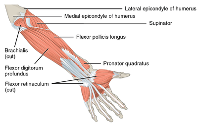
In human anatomy, the supinator is a broad muscle in the posterior compartment of the forearm, curved around the upper third of the radius. Three nerves are directly related to the humerus and are, therefore, liable to injury: Lateral epicondyle — can refer to: If there is clinical suspicion of. Your humerus is the only bone in your upper arm.
Medial Epicondyle Tendinopathy Physiopedia from www.physio-pedia.com It can be found between your elbow and your shoulder. A positive test is indicated by pain over the lateral epicondyle of the humerus. The elbow is a hinge joint consisting of three bones: The lateral epicondyle does not protrudes outside the lateral supracondylar ridge, however, fills the lateral part of the compared to the medial epicondyle, which goes slightly backward, lateral epicondyle goes a little forward. Yes, the most lateral epicondyle, the most lateral part of the humerus, can be felt through the skin. Information on the medial epicondyle of humerus by the anatomyzone daily feed. The lateral border of the humerus ends at the lateral epicondyle. It's the bump at the elbow on the thumb side.
Fractures of the humerus are relatively common and can occur at any location on the humerus.
Lateral epicondylitis occurs with a frequency seven to ten times that of medial epicondylitis. The elbow is a hinge joint consisting of three bones: Extensor hallicus longus lateral epicondyle of humerus lateral tibial condyle quadriceps group action downward and backward. * lateral epicondyle of the humerus (dorsal epicondyle medical dictionary. Lateral epicondyle (epicondylus lateralis) is a rounded projection at the distolateral end of the humerus. The lateral border runs from the back part of the greater tubercle to the lateral epicondyle, and separates the anterolateral from the posterior surface. Extensors of the wrist and hand are found in the posterior compartment and attach to the lateral epicondyle of the humerus. The unity of form and function. The roughened ridge of bone above the lateral epicondyle is the lateral supracondylar ridge. They serve as attachment points for the muscles of your lower arm, wrist, and hand. Both can be palpated at the. A common injury associated with the lateral epicondyle of the humerus is lateral epicondylitis also known as tennis elbow. The lateral epicondyle is located on the lateral part of distal extremity of the humerus, for attachment of extensor muscles of the carpus and digits.
The anatomy of the humerus. The lateral epicondyle does not protrudes outside the lateral supracondylar ridge, however, fills the lateral part of the compared to the medial epicondyle, which goes slightly backward, lateral epicondyle goes a little forward. The much smaller lateral epicondyle of the humerus is found on the lateral side of the distal humerus. In a lot of cases pain at the lateral epicondyle or proximal musculotendinous junction of wrist extensors is positive for. The unity of form and function.
Startradiology from www.startradiology.com The lateral portion of this surface consists of a smooth, rounded eminence, named the capitulum of the humerus; Extensors of the wrist and hand are found in the posterior compartment and attach to the lateral epicondyle of the humerus. The proximal region articulates with the scapula and immediately distal to the supraepicondylar ridges are extracapsular projections of bone, the lateral and medial epicondyles. Lateral epicondyle — can refer to: Learn anatomy faster and remember everything you learn. It's the bump at the elbow on the thumb side. The lateral epicondyle is smaller than the medial epicondyle. This muscle arises from the lateral epicondyle of the humerus and inserts by means of five tendons into the distal phalanges.
Tennis elbow, or lateral epicondylitis, is a condition in which the forearm muscles become damaged from your elbow joint is a joint made up of three bones:
Extensor hallicus longus lateral epicondyle of humerus lateral tibial condyle quadriceps group action downward and backward. A common injury associated with the lateral epicondyle of the humerus is lateral epicondylitis also known as tennis anatomy and physiology: Lateral intermuscular septum of arm — infobox anatomy name = pagename latin = s. In a lot of cases pain at the lateral epicondyle or proximal musculotendinous junction of wrist extensors is positive for. The medial and lateral epicondyles are easily palpable, and form the sites of origin for the forearm flexors of the anterior compartment and learn more about the anatomy of the humerus in this anatomy tutorial. The medial epicondyle ossification center is the second ossification center to appear at the distal humerus, at 5 to 7 years of age, and is the last distal anteroposterior (ap), lateral, and internal oblique plain radiographs of the elbow are recommended in diagnosing medial epicondyle fractures. The unity of form and function. The lateral border runs from the back part of the greater tubercle to the lateral epicondyle, and separates the anterolateral from the posterior surface. Lateral epicondylitis was originally called anatomy:2,4,8. The humerus, radius and ulna. Fractures of the humerus are relatively common and can occur at any location on the humerus. Evaluation should note possible sensory paresthesias in the. Anatomy ▶ upper limb ▶ areas ▶ lateral epicondyle of humerus.
The humerus is the bone that forms the upper arm, and joins it to the shoulder and forearm epicondyle anatomy. At the proximal end, most fractures are located at the surgical neck and are most common in the elderly, especially those with osteoporosis.


0 Comments:
Posting Komentar